1、Cell:线虫中全脑神经元鉴定的多色图谱
2020年12月29日,来自美国哥伦比亚大学Eviatar Yemini等研究人员在期刊《细胞》上发表了题为“NeuroPAL: A Multicolor Atlas for Whole-Brain Neuronal Identification in C. elegans.”的研究论文,绘制出线虫中全脑神经元鉴定的多色图谱。
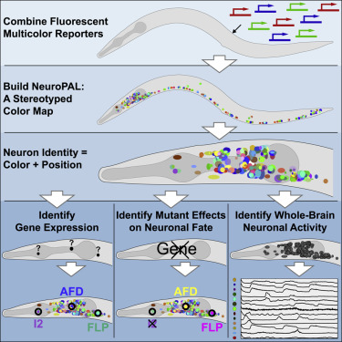
Fig 1|来源cell
研究人员通过设计一种称为NeuroPAL(神经元多色图谱)的多色转基因技术,并在秀丽隐杆线虫中实现了这一目标。NeuroPAL线虫在整个雌雄同体的神经系统中共享一个固定的多色荧光图谱,从而可解析所有的神经元身份。标记有NeuroPAL的神经元在绿色、青色或黄色发射通道中不发射荧光,从而使这个转基因可与基因表达或神经元动力学的许多已报道分子一起使用。
研究人员利用NeuroPAL进行了神经系统范围内神经元鉴定的三种应用。首先,研究人员确定了乙酰胆碱、GABA和谷氨酸的所有代谢型受体的全脑表达模式,从而完成了该交流网络的图谱。其次,研究人员发现了转录因子突变引起的细胞命运变化。第三,研究人员记录了响应吸引和排斥化学感觉线索时的全脑活动,从而表征这些刺激的多模式编码。
Summary
Comprehensively resolving neuronal identities in whole-brain images is a major challenge. We achieve this in C. elegans by engineering a multicolor transgene called NeuroPAL (a neuronal polychromatic atlas of landmarks). NeuroPAL worms share a stereotypical multicolor fluorescence map for the entire hermaphrodite nervous system that resolves all neuronal identities. Neurons labeled with NeuroPAL do not exhibit fluorescence in the green, cyan, or yellow emission channels, allowing the transgene to be used with numerous reporters of gene expression or neuronal dynamics. We showcase three applications that leverage NeuroPAL for nervous-system-wide neuronal identification. First, we determine the brainwide expression patterns of all metabotropic receptors for acetylcholine, GABA, and glutamate, completing a map of this communication network. Second, we uncover changes in cell fate caused by transcription factor mutations. Third, we record brainwide activity in response to attractive and repulsive chemosensory cues, characterizing multimodal coding for these stimuli.
(评论:全面解析全脑图像中的神经元身份是一项重大挑战。)
文章来源:
Eviatar Yemini, Albert Lin et al, NeuroPAL: A Multicolor Atlas for Whole-Brain Neuronal Identification in C. elegans, DOI: 10.1016/j.cell.2020.12.012, Cell:最新IF:36.216
2、Nature Genetics:破译影响人类”颜值”的遗传因素
2020年12月7日,来自比利时鲁汶大学Peter Claes等研究人员在《自然—遗传学》杂志上发表了题为“Insights into the genetic architecture of the human face.”的研究结果,发现破译影响人类面部形成的遗传因素。
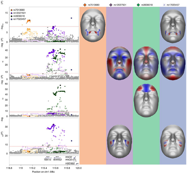
Fig 2|TBX15-WARS2 multi-peak locus.(来源:Nature Genetics)
研究人员表示,Achley和Hall在1991年概括了当代生物学的主要问题之一:在发育过程中如何出现复杂的形态结构以及在进化过程中如何改变形态。这个问题继续困扰着生物学家、遗传学家、人类学家和临床医生近三十年。人脸就是这样一种典型的复杂形态结构,且高度多态,是遗传、细胞和环境因素复杂协调的结果。通过先前的全基因组关联研究(GWAS),已激发下100多个基因座参与正常范围的面部形态塑造,但综合表征影响人脸的遗传结构仍然具有挑战性。
使用多变量全基因组关联研究对8,246例欧洲个体进行荟萃分析,研究人员确定了203个与正常范围面部变异相关的全基因组信号(120个在研究范围内也有意义)。后续分析表明,围绕这些信号的区域在颅神经crest细胞和颅面组织中的增强子活性得到了富集,数个区域藏有多种信号,它们与不同的面部表型相关,并且有证据表明这些变异可能具有协同作用。总而言之,这些分析为了解个体和协调的遗传行为如何塑造复杂的形态特征提供了见识。
Summary
The human face is complex and multipartite, and characterization of its genetic architecture remains challenging. Using a multivariate genome-wide association study meta-analysis of 8,246 European individuals, we identified 203 genome-wide-significant signals (120 also study-wide significant) associated with normal-range facial variation. Follow-up analyses indicate that the regions surrounding these signals are enriched for enhancer activity in cranial neural crest cells and craniofacial tissues, several regions harbor multiple signals with associations to different facial phenotypes, and there is evidence for potential coordinated actions of variants. In summary, our analyses provide insights into the understanding of how complex morphological traits are shaped by both individual and coordinated genetic actions.
(评论:这项研究不仅可以增进我们对人脸遗传学的理解,而且还有助于我们更好地了解人类面部先天缺陷的形成因素。)
文章来源:
Julie D. White, Karlijne Indencleef, Insights into the genetic architecture of the human face. DOI: 10.1038/s41588-020-00741-7, Nature Genetics:最新IF:25.455
3、Science:揭示preTCR与其配体配对方式
2020年12月17日,来自美国达纳-法伯癌症研究所Ellis L. Reinherz和Jia-huai Wang研究组合作在《科学》杂志上发表了题为“Pre–T cell receptors topologically sample self-ligands during thymocyte β-selection.”的研究结果,发现在胸腺细胞β-选择过程中,pre-T细胞受体(preTCRs)以拓扑结构方式采样自身配体。
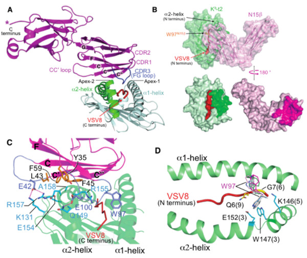
Fig 3| reTCRβ-pΜΗC-I复合物结构、配位分子(来源science)
使用X射线晶体学,他们显示了preTCR如何将其单个可变域(Vβ)的凹面β-折叠表面“水平”地抓住凸出的MHCα2-螺旋。相比之下,αβTCR的目的是使其配对的VαVβ模块的所有六个互补决定区(CDR)环识别“垂直”头对头结合中与MHC分子(pMHCs)结合的肽。preTCR的拓扑拟合可确保CDR3β到达肽的特征性C末端片段以进行pMHC采样,从而建立随后的αβTCR典型对接模型。
“水平”对接排除了种系CDR1β–和CDR2β–MHC的结合,从而在αβTCR介导的选择优化之前拓宽了β链库的多样性。因此,一个亚基相继调节相关多组分受体的识别逻辑。
据悉,自我歧视是在胸腺细胞发育过程中编程的一个关键但不确定的分子过程,它需要大量的preTCRs和αβTCR。
Summary
Self-discrimination, a critical but ill-defined molecular process programmed during thymocyte development, requires myriad pre-T cell receptors (preTCRs) and αβTCRs. Using X-ray crystallography, we show how a preTCR applies the concave β-sheet surface of its single variable domain (Vβ) to “horizontally” grab the protruding MHC α2-helix. By contrast, αβTCRs purpose all six complementary-determining region (CDR) loops of their paired VαVβ module to recognize peptides bound to MHC molecules (pMHCs) in “vertical” head-to-head binding. The preTCR topological fit ensures that CDR3β reaches the peptide’s featured C-terminal segment for pMHC sampling, establishing the subsequent αβTCR canonical docking mode. “Horizontal” docking precludes germline CDR1β– and CDR2β–MHC binding to broaden β-chain repertoire diversification before αβTCR-mediated selection refinement. Thus, one subunit successively attunes the recognition logic of related multicomponent receptors.
(评论:认识了可视化的preTCR-pΜΗC复合物晶体结构,让大家也了解到preTCR的拓扑结构与CDR3在“TCR-β选择”中重要作用,为研究preTCR介导的T细胞分化发育的科学工作者,提供了一个新思路。)
文章来源:
Xiaolong Li, Réka Mizsei et al, Pre–T cell receptors topologically sample self-ligands during thymocyte β-selection. DOI: 10.1126/science.abe0918, 最新IF:41.037